
Fungal Nail Infection (Onychomycosis) – Causes, Types & Treatment
What Is a Fungal Nail Infection?
A fungal nail infection, clinically termed onychomycosis, is a progressive fungal disease affecting the nail unit, which includes the nail plate, nail bed, and surrounding cuticle tissue. It commonly affects toenails, though fingernails can also be involved.
The infection occurs when fungi—particularly dermatophytes such as Trichophyton rubrum, along with yeasts (e.g. Candida) and non-dermatophyte moulds—penetrate the nail structure, leading to discolouration, thickening, brittleness, and, in advanced cases, nail detachment.
These organisms thrive in warm, moist environments such as locker rooms, communal showers, and closed footwear. Without proper diagnosis and management, onychomycosis can persist for years, impacting both nail health and foot hygiene.
Onychomycosis is one of the most prevalent nail disorders treated by podiatrists, accounting for over 50% of all nail-related consultations.
Medically reviewed by: Dr Priya Anand DPM. Last update: 18th May 2026
Book your appointment
Onychomycosis vs Nail Fungus vs Tinea Unguium
These three terms often appear interchangeably, but they carry subtle distinctions important in clinical practice:
| Term | Definition | Clinical Usage |
|---|---|---|
| Onychomycosis | Umbrella medical term for any fungal infection of the nail | Diagnostic codes, medical literature |
| Fungal nail infection | Common language term for onychomycosis | Patient-facing education and communications |
| Tinea unguium | Subtype of onychomycosis specifically caused by dermatophytes | Mycology, dermatology, lab reports |
Understanding this terminology helps patients recognise diagnostic accuracy and treatment relevance.
How Common Is It?
Fungal nail infections are highly prevalent, particularly among the following risk groups:
- Adults aged 50 years and older
- Individuals with diabetes, vascular disease, or immunosuppression
- People with recurrent athlete’s foot (tinea pedis)
- Gym users or swimmers exposed to damp, shared environments
Epidemiology:
- ~10% of the general population is affected by onychomycosis
- Prevalence increases to 25–30% in adults over 60
- Over 50% of nail disorders seen in clinical podiatry are fungal in origin
Fungal nail infections represent the majority of nail disorders treated clinically. According to the British Association of Dermatologists, over half of all nail disease cases assessed in dermatology or podiatry settings are attributable to onychomycosis.
The infection is contagious, spreading via:
- Shared footwear, towels, and nail tools
- Direct skin-to-skin contact
- Communal areas like spas, gyms, swimming pools
- Self-inoculation from adjacent skin conditions like tinea pedis

What Causes Fungal Nail Infections?
Primary Fungal Organisms
Fungal nail infections (onychomycosis) are primarily caused by dermatophytes—a group of fungi that feed on keratin found in nails, skin, and hair. These pathogens penetrate the nail plate or nail bed through small cracks or trauma, especially when conditions are warm, moist, and enclosed.
Clinical Classification of Fungi Involved in Onychomycosis:
| Fungal Type | Common Species | Typical Context |
|---|---|---|
| Dermatophytes | Trichophyton rubrum, T. interdigitale | Most common in toenail infections |
| Yeasts (Candida spp.) | Candida albicans | More common in fingernails, especially in women and immunocompromised individuals |
| Non-Dermatophyte Moulds | Scopulariopsis brevicaulis, Fusarium spp. | Often found in soil, associated with nail trauma |
Dermatophytes are responsible for over 90% of fungal toenail infections in the UK.
Risk Factors for Onychomycosis
Multiple intrinsic and environmental risk factors increase susceptibility to fungal nail infections.
Health & Medical Predisposition
- Diabetes mellitus: Impaired circulation and reduced immunity
- Peripheral vascular disease: Poor blood flow to extremities
- Immune suppression: From chemotherapy, HIV, or corticosteroids
- History of tinea pedis (athlete’s foot): Often a precursor to nail infection
Nail-Related Triggers
- Nail trauma: Repeated pressure, running, or poorly fitted footwear
- Improper nail care: Over-clipping or using contaminated instruments
- Nail psoriasis or eczema: Compromises the nail’s protective barrier
Environmental Exposure
- Communal showers or changing rooms: High-risk zones for dermatophyte transmission
- Unsterilised nail salons: Risk of Candida or mould contamination
- Wearing occlusive shoes: Creates a damp environment ideal for fungal growth
How Fungal Nail Infections Spread
Onychomycosis is contagious and may spread via:
- Direct contact with infected skin or nails (e.g. tinea pedis)
- Shared personal items like towels, socks, or footwear
- Nail salons with inadequate sterilisation practices
- Public surfaces such as locker room floors or swimming pool decks
A podiatrist will often trace infection origin through microtrauma, nail fold breakdown, or pre-existing fungal skin conditions.
Types of Fungal Nail Infections
Onychomycosis, the medical term for fungal nail infection, comprises several clinically distinct subtypes. These are defined by their point of fungal entry, pathogen type, and progression pattern within the nail unit. Proper classification helps podiatrists determine the most effective treatment modality, whether topical, systemic, or laser-based.
Subtypes of Onychomycosis
| Subtype | Description | Common Causative Fungi |
|---|---|---|
| Distal Lateral Subungual Onychomycosis (DLSO) | Begins at the edge or side of the nail and progresses under the plate | Trichophyton rubrum |
| White Superficial Onychomycosis (WSO) | Surface-level infection with chalky white patches | T. mentagrophytes |
| Proximal Subungual Onychomycosis (PSO) | Starts near the cuticle and migrates outward | Associated with immunodeficiency |
| Endonyx Onychomycosis | Affects the internal nail plate without lifting | Rare, often dermatophyte-driven |
| Total Dystrophic Onychomycosis (TDO) | Complete nail destruction from chronic infection | End-stage of untreated DLSO or PSO |
| Candida Onychomycosis | Affects fingernails more than toenails; often with swelling and pain | Candida albicans |
DLSO – Distal Lateral Subungual Onychomycosis
DLSO is the most common form of fungal nail infection, accounting for over 80–90% of clinical cases.
- Fungal entry occurs through the distal or lateral nail bed.
- Early signs include yellow discoloration, subungual debris, and thickened nail plates.
- Often associated with chronic tinea pedis (athlete’s foot) and tight footwear.
Podiatrists typically identify DLSO through nail examination and KOH microscopy.

WSO – White Superficial Onychomycosis
White superficial onychomycosis affects the nail surface directly:
- Presents as powdery, white lesions on the nail plate
- More common in younger patients
- Can be scraped off in early stages
- Responds well to topical antifungal lacquers
PSO – Proximal Subungual Onychomycosis
Proximal subungual onychomycosis begins at the cuticle or proximal nail fold, progressing outward.
- May indicate immunosuppression, such as HIV or chemotherapy
- Often misdiagnosed as nail trauma or psoriasis
- Requires laboratory confirmation and potentially systemic treatment
PSO is considered a marker condition in immunocompromised patients.
Endonyx Onychomycosis
This rare form involves fungal invasion of the nail plate without nail separation or debris.
- Nail may appear milky or discoloured, but not lifted
- Common in tropical regions
- Typically caused by Trichophyton soudanense or T. violaceum
TDO – Total Dystrophic Onychomycosis
This represents end-stage onychomycosis, often due to long-standing, untreated fungal invasion.
- Nail plate is completely destroyed or disintegrated
- Accompanied by pain, secondary infections, and functional issues
- May necessitate nail avulsion or laser therapy as a salvage approach
Candida Onychomycosis
Caused by yeasts rather than dermatophytes, Candida onychomycosis is more frequent in:
- Fingernails, especially among healthcare workers or those with chronic wet hands
- People with diabetes, thyroid disorders, or poor peripheral circulation
Often presents with paronychia (red, swollen nail folds) and yellow-brown discoloration.
Candida onychomycosis is primarily a yeast-driven infection, often affecting fingernails rather than toenails. A systematic review in the Journal of Fungi reports that Candida albicans and other yeasts are strongly linked with paronychia and periungual inflammation, particularly in individuals with frequent water exposure or chronic illnesses such as diabetes.
Signs and Symptoms of Fungal Nail Infections
Early Clinical Indicators
Onychomycosis often begins with subtle, painless changes that are easy to dismiss. However, early recognition allows for more effective and less invasive treatment.
Key early signs include:
- Yellow or white discolouration starting at the nail edge
- Chalky, powdery patches (particularly in white superficial onychomycosis)
- Loss of natural shine or clarity in the nail plate
- Mild thickening or changes in nail surface texture
- Brittle or frayed nail edges
Distal Lateral Subungual Onychomycosis (DLSO) often begins at the nail’s free edge and progresses beneath the plate.
Progression and Severity Markers
As the infection advances, fungal colonisation spreads deeper, triggering structural and functional deterioration of the nail.
Symptoms of moderate to severe onychomycosis:
- Subungual hyperkeratosis – thickening under the nail plate
- Onycholysis – the nail separating from its bed
- Irregular nail shape or full deformation
- Discolouration deepens to yellow, brown, or green
- Foul odour from fungal metabolic byproducts
- Pain or pressure when walking or wearing shoes
In Total Dystrophic Onychomycosis (TDO), the entire nail may disintegrate, necessitating full debridement or surgical management.
Peripheral and Soft Tissue Symptoms
While fungal nail infection primarily affects the nail unit, associated signs in surrounding tissue can indicate complication or spread:
- Paronychia – inflammation and redness around the nail folds
- Peeling or scaling skin between toes (coexistent tinea pedis)
- Recurring ingrown toenails due to thickened or distorted nails
- Skin fissures or ulceration in patients with diabetes or neuropathy
These signs are especially relevant in immunocompromised individuals, where even minor infections can escalate rapidly.
Symptom Overlap and Differential Diagnosis
Not all nail abnormalities are fungal. Several conditions mimic onychomycosis, making professional diagnosis essential.
| Symptom | Fungal Nail Infection | Nail Psoriasis | Nail Trauma |
|---|---|---|---|
| Discolouration | Yellow, green, or white | Oil-drop spots | Reddish-brown bruising |
| Nail thickening | Common (hyperkeratosis) | Possible | Possible |
| Onycholysis | Yes, progresses with severity | Yes | Yes, sudden onset |
| Pain | With footwear or pressure | Rare | Sharp or transient |
| Nail pitting | Rare | Frequent | Absent |
A podiatrist will typically perform a fungal nail test (mycological culture or KOH prep) to confirm diagnosis.
Diagnosing Fungal Nail Infections
A correct diagnosis of onychomycosis — the clinical term for fungal nail infection — is essential before starting treatment. Many non-fungal nail disorders such as psoriasis, eczema, or trauma can mimic fungal infection, leading to inappropriate or ineffective therapy when misdiagnosed.
Visual Examination by a Podiatrist
Your diagnosis journey typically begins with a consultation with a HCPC-registered podiatrist. Using clinical expertise, they will assess:
- Nail colour, including signs of yellow, green, or brown discolouration
- Nail thickness and shape, checking for subungual hyperkeratosis
- Presence of onycholysis – detachment of the nail from the nail bed
- Texture, debris, or brittle appearance
- Surrounding skin for paronychia, scaling, or signs of tinea pedis
A podiatrist’s role includes differential diagnosis — distinguishing true fungal infections from similar nail pathologies.
Laboratory Confirmation Methods
A visual diagnosis alone is not enough. Mycological confirmation using lab testing is strongly recommended, particularly before initiating laser or systemic antifungal treatments.
1. Fungal Nail Test (Nail Clipping & Culture)
- A section of the nail is clipped and sent for fungal culture
- Confirms the presence and type of fungus, e.g. Trichophyton rubrum
- Results returned in 2–3 weeks
- Often required for private laser clinics or GPs prescribing oral antifungals
2. KOH Microscopy (Potassium Hydroxide Test)
- A quick diagnostic method using nail scrapings
- KOH dissolves keratin, revealing fungal elements (hyphae) under a microscope
- Offers same-day preliminary results
- Frequently used in in-clinic podiatry settings
3. PCR Fungal DNA Testing (Private Clinics Only)
- Detects fungal DNA at high sensitivity
- Useful for unclear cases or failed cultures
- Ideal for immunocompromised patients or where rapid confirmation is required
When Diagnostic Testing Is Recommended
Fungal nail testing should be performed when:
- Starting Erchonia® Lunula Laser therapy
- Prescribing oral antifungal medications (e.g. terbinafine, itraconazole)
- Previous treatment has failed
- Symptoms are atypical or only affecting a single nail
- Patient has underlying conditions such as diabetes or vascular disease
Testing also helps differentiate onychomycosis from non-fungal nail dystrophies.
Comparing Diagnostic Accuracy
| Method | Turnaround | Accuracy | Clinical Notes |
|---|---|---|---|
| KOH microscopy | Same-day | Moderate | Quick, but may miss non-viable fungi |
| Fungal culture | 2–3 weeks | High | Gold standard; identifies fungal species |
| PCR DNA test | 2–5 days | Very high | Expensive; private labs only |
PCR offers high diagnostic yield but may not differentiate viable from non-viable organisms.
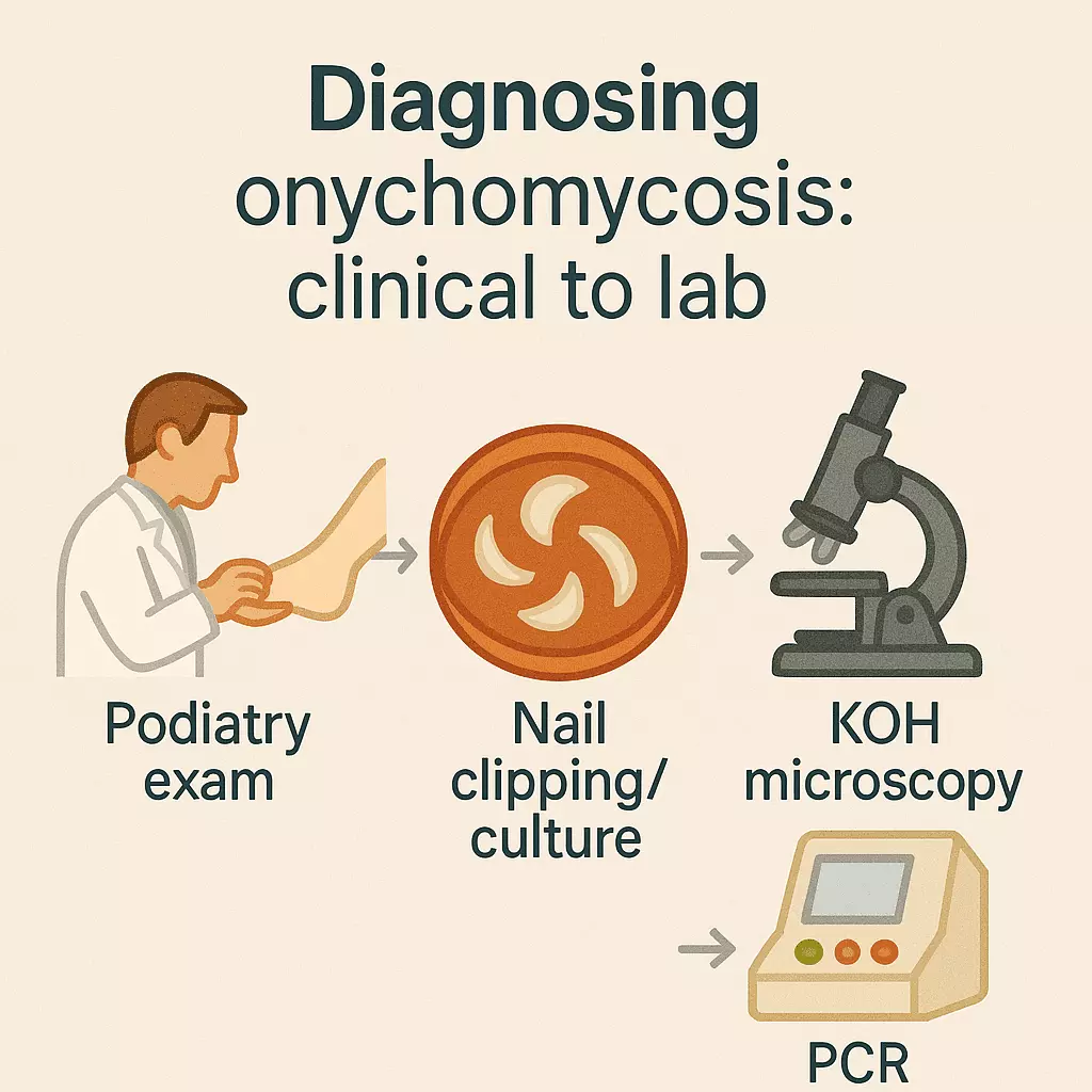
Diagnostic workflow and instruments are covered in our foot conditions hub, including testing pathways for nails, skin, and biomechanical issues.
Treatment Options for Fungal Nail Infections
Effectively treating onychomycosis — a fungal infection of the toenails or fingernails — requires selecting the right therapy based on infection type, severity, and patient suitability. Treatment should always follow confirmed diagnosis by a podiatrist to ensure accuracy and avoid mistreating non-fungal conditions.
Topical Antifungal Therapies
Topical antifungal medications are ideal for mild infections, especially white superficial onychomycosis (WSO) or early-stage distal lateral subungual onychomycosis (DLSO).
Common Topical Agents:
- Amorolfine 5% nail lacquer – applied weekly, often for 6–12 months
- Ciclopirox (Penlac) – daily use; requires nail debridement for best results
- Natural options (e.g., tea tree oil) – some anecdotal benefit, limited clinical evidence
Efficacy depends on consistent application, thorough debridement, and patience — visible improvement can take 9–12 months.
Systemic Oral Medications
Oral antifungals are suited for moderate to severe onychomycosis involving nail matrix or full-thickness infection.
First-line Drugs:
- Terbinafine (Lamisil) – typically 250 mg daily for 6–12 weeks
- Itraconazole (Sporanox) – pulsed or continuous dosing
Advantages:
- Higher cure rates than topical agents
- Targets fungal reservoirs in the skin and nails
Considerations:
- May cause liver enzyme elevation — LFTs (liver function tests) are often required
- Interacts with several medications
- Contraindicated in pregnancy, liver disease, or certain cardiovascular conditions
Oral therapy should only be prescribed after a confirmed fungal nail test (mycology or PCR).
Laser Therapy (Erchonia® Lunula Laser)
The Erchonia® Lunula Laser is a cutting-edge, low-level laser therapy (LLLT) designed to eradicate fungal pathogens without pain, heat, or side effects.
Key Features:
- Uses dual-wavelength low-level laser light (405nm and 635nm)
- Stimulates immune response and disrupts fungal cell metabolism
- Entirely non-invasive and safe for all ages, including diabetics
Treatment Protocol:
- Usually involves 4 weekly sessions
- No downtime or recovery required
- Often used in conjunction with nail debridement for maximum effect
Freedom Clinics offers advanced Fungal Nail Laser Treatment with Erchonia® technology — a safe choice for patients who cannot tolerate systemic medications.
Nail Debridement (Clinical Support Procedure)
Debridement involves the mechanical removal of:
- Infected keratin and fungal debris
- Thickened or distorted nail layers
Performed by a podiatrist using sterile tools, this:
- Reduces fungal load
- Enhances topical agent penetration
- Alleviates discomfort or pressure from hyperkeratotic nails
Often recommended alongside topical, laser, or oral therapies for synergistic benefit.
Hygiene & At-Home Maintenance
Long-term success in fungal nail treatment depends heavily on home hygiene and preventive care:
- Keep feet dry and nails trimmed
- Disinfect shoes and socks with antifungal sprays
- Avoid walking barefoot in communal areas
- Don’t share nail clippers or foot files
- Use medicated foot powders if prone to tinea pedis (athlete’s foot)
Recurrence is common without lifestyle changes and follow-up care.
Choosing the Right Treatment Path
| Clinical Factor | Recommended Approach |
|---|---|
| Mild, superficial infection | Topical treatment ± debridement |
| Moderate DLSO | Oral antifungals or laser therapy |
| Contraindicated for oral meds | Laser or topical with podiatry support |
| Severe nail dystrophy (TDO) | Debridement, possible nail removal, laser as adjunct |
| High-risk patient (diabetes, liver disease) | Laser therapy under clinical supervision |
A customised treatment plan, guided by a podiatrist and based on confirmed diagnosis, offers the best chance of full recovery.

Prevention & Aftercare for Fungal Nail Infections
Successfully treating onychomycosis is only half the journey. The other half is preventing recurrence — a common challenge if foot hygiene, footwear, and nail environments are not properly managed. Reinfection typically stems from fungal spores lingering in shoes, socks, or shared surfaces.
Daily Foot Hygiene & Moisture Control
Moisture is the enemy of healthy toenails. Creating a dry, clean environment disrupts fungal growth and reduces the risk of relapse.
Best Practices:
- Dry feet thoroughly, especially between the toes, after showers or exercise
- Use antifungal foot powder daily (e.g. tolnaftate-based formulations)
- Wear moisture-wicking socks and breathable shoes
- Avoid prolonged dampness — change socks if your feet get sweaty
- Never share towels, socks, or slippers
Consistency is key. Fungi flourish in warm, moist conditions — eliminating these drastically lowers the chance of regrowth.
Disinfecting Footwear & Surfaces
Even after the nail appears clear, fungal spores can persist inside shoes and socks.
Recommended Measures:
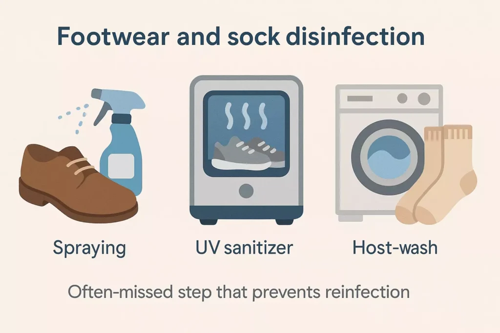
- Spray inside shoes with antifungal shoe disinfectant
- Use UV light sanitisers to neutralise spores in trainers or insoles
- Wash socks at 60°C or higher
- Replace heavily worn shoes or slippers used during the infection period
- Clean shared surfaces like floors or mats if barefoot use occurred
Proper footwear hygiene is often overlooked — yet it’s a leading factor in fungal reinfection.
Routine Podiatrist Follow-Up
Following treatment, it’s essential to continue monitoring your nail health. A podiatrist will assess:
- Regrowth of healthy nail tissue
- Any signs of subclinical recurrence
- Whether further nail debridement is needed
- Suitability for prophylactic topical therapy
Follow-up schedule:
Every 3–6 months, especially for high-risk groups such as:
- Diabetics
- Elderly individuals
- Athletes and swimmers
- Immunocompromised patients
Your podiatrist acts as your partner in long-term foot health — early detection means faster action.
Preventive Topical Therapy (Prophylaxis)
Once the infection is cleared, maintenance therapy is a proactive way to keep onychomycosis from returning.
Common Options:
- Amorolfine lacquer applied weekly
- Tea tree oil as a natural barrier (limited clinical data)
- Tolnaftate or miconazole powders/sprays inside shoes
- Topical antifungal creams around toes and nail folds
Weekly application post-treatment, particularly after laser or oral therapy, is highly effective for preventing fungal re-establishment.
Lifestyle Habits to Reduce Reinfection Risk
Changing small behaviours makes a big difference:
| Habit | Why It Matters |
|---|---|
| Change socks daily (or twice if needed) | Controls moisture |
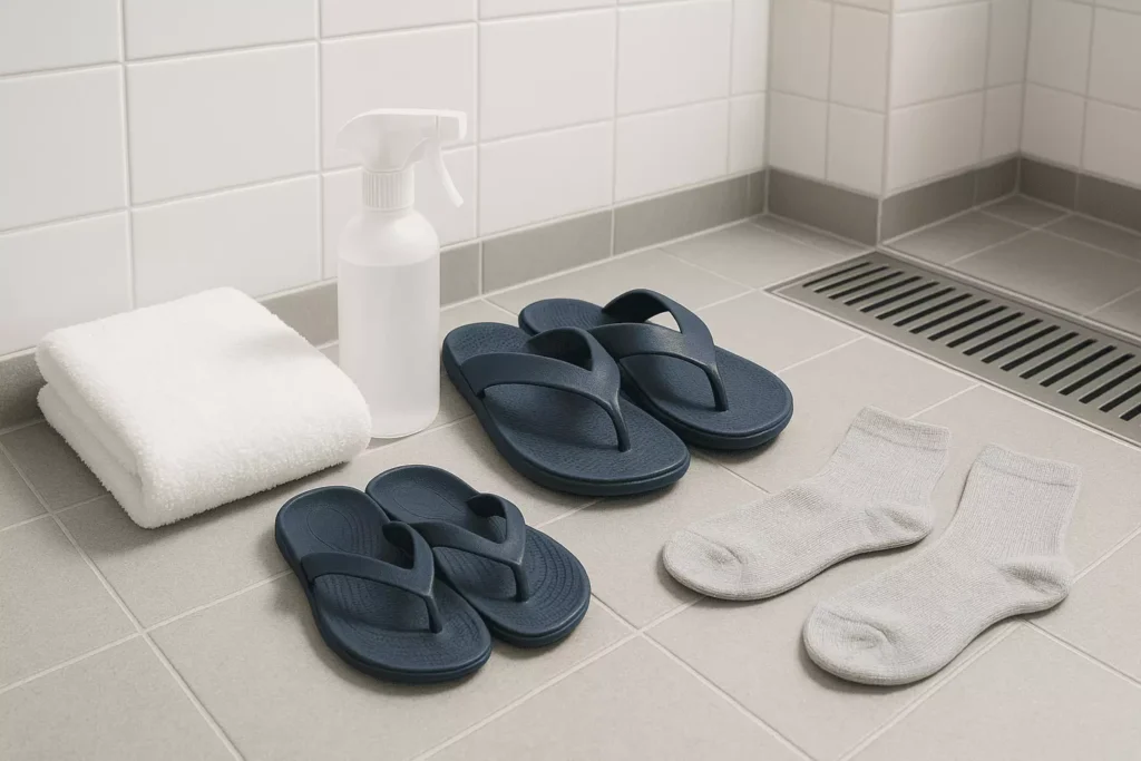
| Wear sandals in communal showers | Prevents surface transmission |
| Disinfect nail tools after each use | Avoids spore transfer |
| Avoid sharing pedicure instruments | Reduces cross-contamination |
| Alternate between two pairs of shoes | Allows full drying time |
Relapse often occurs not from treatment failure — but from re-exposure to fungal environments.
For ongoing care, clinic-based maintenance, and follow-up schedules, explore our Podiatry & Chiropody services.
Related Conditions
- Tarsal tunnel syndrome: tibial nerve entrapment overview
- Hallux rigidus (stiff big toe) clinical guide
- Posterior tibial tendon dysfunction (PTTD) management
- Sesamoiditis pain and load modification
- Rheumatoid arthritis in the feet: podiatry care
- Peripheral neuropathy and foot protection
- Osteoarthritis of the foot & ankle
- Foot drop: causes and support options
- Charcot foot (neuroarthropathy) essentials
- Gout in the foot: flare management
- Achilles tendonitis: symptoms and treatment
- Bunions (hallux valgus) treatment options
- Lisfranc injuries: midfoot stability
- Diabetic foot ulcers: prevention & care
- Morton’s neuroma (forefoot nerve pain)
- Corns and calluses: debridement & offloading
- Baxter’s nerve entrapment (heel pain)
- Plantar fibroma: fascia nodules & care
Our Podiatrists & Chiropodists


References
- M. Ameen, J.T. Lear, V. Madan, M.F. Mohd Mustapa, M. Richardson, J.R. Hughes, A. Sahota, M. Griffiths, A.J. McDonagh, S. Punjabi, D.A. Buckley, I. Nasr, V.J. Swale, C.E. Duarte Williamson, P.M. McHenry, N.J. Levell, T. Leslie, E. Mallon, K. Towers, R. Davis, C. Saunders, S.E. Haveron, L.S. Exton, British Association of Dermatologists’ guidelines for the management of onychomycosis 2014, British Journal of Dermatology, Volume 171, Issue 5, 1 November 2014, Pages 937–958, https://doi.org/10.1111/bjd.13358
- Zisova, L., & Valtchev, V. (2021). Candida Onychomycosis: Clinical Features and Treatment. Journal of Fungi, 7(12), 1065. https://doi.org/10.3390/jof7121065

