
Posterior Tibial Tendon Dysfunction (PTTD)
Posterior Tibial Tendon Dysfunction (PTTD) is a degenerative condition of the posterior tibial tendon—a key structure that supports the medial longitudinal arch and helps control foot movement during walking. When this tendon becomes inflamed, weakened, or torn, it loses its ability to stabilise the arch, often leading to adult-acquired flatfoot, gait instability, and persistent medial ankle pain.
In the early stages, PTTD may feel like a nagging soreness or sharp foot pain on the inside of the ankle, especially after prolonged standing or walking. As the tendon dysfunction progresses, it causes visible arch collapse, overpronation (inward rolling of the foot), and increasing difficulty with walking or maintaining balance. Without timely intervention, PTTD can advance to rigid flatfoot, ankle arthritis, and compensatory pain in the knees, hips, or lower back due to altered gait mechanics.
At Freedom Clinics, located in the heart of Canary Wharf, London, we specialise in the diagnosis and treatment of posterior tibial tendon dysfunction. Our experienced podiatry team provides custom orthotic solutions, biomechanical assessments, and stage-specific treatment plans tailored to restore function and prevent progression. Whether you’re experiencing arch pain, flat feet, or suspect tendon damage, our London-based clinicians can help you move with confidence again.
Medically reviewed by: Dr Priya Anand DPM. Last update: 18th May 2026
Book your appointment
Understanding Posterior Tibial Tendon Dysfunction
Anatomical Role of the Posterior Tibial Tendon
The posterior tibial tendon is one of the most important stabilising tendons in the lower limb. Originating from the posterior tibialis muscle in the calf, it runs behind the inner ankle and inserts into several bones on the medial side of the foot, including the navicular. Its main function is to support the medial longitudinal arch and to invert the foot during the stance phase of the gait cycle.
This tendon plays a crucial role in controlling overpronation and distributing weight efficiently during walking and running. When functioning properly, it acts as a dynamic structure that prevents the arch from collapsing. However, due to its high workload and critical biomechanical role, it is susceptible to overuse, inflammation, and degeneration, especially in adults over 40 or those with abnormal gait mechanics.
A study at the Royal National Orthopaedic Hospital reported that symptomatic posterior tibial tendon dysfunction affects approximately 3.3% of women over age 40, indicating a predisposition of this tendon to overuse and degeneration in that age group.
What Leads to Posterior Tibial Tendon Dysfunction?
Posterior Tibial Tendon Dysfunction (PTTD) typically begins with tendinitis, an inflammatory response to overuse, repetitive strain, or acute injury. This initial phase is marked by pain on the inside of the ankle, mild swelling, and tenderness during activity. As the dysfunction progresses, the tendon becomes elongated or partially torn, compromising its ability to support the foot’s arch.

The weakened tendon loses tension, resulting in posterior tibial tendon insufficiency. This leads to a progressive collapse of the medial arch, reduced foot stability, and impaired control over foot positioning during gait. Without intervention, the condition often transitions into chronic flatfoot deformity, with secondary effects on the knees, hips, and even lower back due to altered weight distribution.
A systematic review published in Journal of Foot and Ankle Research by the University of Queensland team showed that individuals with PTTD have significantly lower arch height and poorer heel-rise endurance, confirming the progressive collapse of the medial arch and impaired foot control.
How PTTD Causes Flatfoot and Gait Instability
When the posterior tibial tendon fails, the medial longitudinal arch no longer receives adequate support. This causes the foot to roll inward excessively—a condition known as overpronation—and leads to adult-acquired flatfoot deformity. This collapse alters the biomechanics of walking, making it harder for patients to maintain balance or bear weight evenly.
Over time, patients with untreated PTTD may develop:
- A visibly flattened foot shape
- Outward deviation of the heel (hindfoot valgus)
- Inward collapse of the ankle (medial bulging)
- Increased stress on the tibia and hip joints
Unlike congenital flatfoot, PTTD-induced flatfoot is acquired later in life and is directly linked to the failure of the posterior tibial tendon. While the condition is progressive, it is often reversible or manageable if diagnosed early and treated with orthotic support and targeted rehabilitation.
Signs and Symptoms of Posterior Tibial Tendon Dysfunction
Identifying the symptoms of Posterior Tibial Tendon Dysfunction (PTTD) early is crucial for effective treatment. As the posterior tibial tendon weakens or fails, the foot’s structural integrity begins to decline, leading to progressive medial ankle pain, arch collapse, and gait instability. These symptoms are classified by clinical stage, each reflecting the severity of tendon dysfunction and associated foot deformity.
Early-Stage Symptoms of PTTD (Stage I)
In Stage I, the condition presents as posterior tibial tendinitis—an early inflammatory response without structural tearing. The foot retains its normal shape, but the tendon’s function begins to degrade.
Common signs:
- Pain and swelling along the inner ankle, particularly behind the medial malleolus
- Discomfort after long walks, runs, or prolonged standing
- Mild fatigue in the posterior tibialis muscle
- Localised tenderness when pressing the medial longitudinal arch
- Normal foot alignment when not weight-bearing
At this reversible stage, early intervention with orthotics, rest, and strengthening can prevent long-term damage.
Progressive Symptoms and Functional Changes (Stage II)
Stage II marks the onset of posterior tibial tendon insufficiency, where the tendon has stretched or partially torn. The arch begins to collapse, and functional symptoms become more pronounced.
Key indicators:
- Visible flattening of the medial arch (adult-acquired flatfoot)
- Overpronation—inward rolling of the ankle during gait
- Pain during stair climbing or toe-off
- Difficulty performing a single-leg heel raise
- “Too many toes” sign: lateral toes are visible when viewed from behind due to forefoot abduction
- Tenderness along the tendon pathway
This stage is typically when patients first notice visible changes in foot shape and experience reduced balance or walking confidence.
Structural Collapse and Joint Involvement (Stages III–IV)
In Stage III, the condition becomes rigid and structurally fixed. By Stage IV, joint degeneration extends into the ankle.
Advanced symptoms include:
- Rigid flatfoot deformity with no flexibility
- Hindfoot valgus — outward tilting of the heel bone
- Pain along the subtalar joint and lateral foot
- Difficulty standing or walking without orthotic bracing or support
- Pain in knees, hips, or lower back due to altered biomechanics
- Signs of ankle arthritis — joint stiffness, swelling, and reduced motion
Once rigidity and arthritis occur, conservative treatments become less effective, and surgical intervention may be necessary to correct deformities and relieve pain.
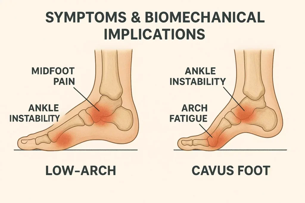
Stages of Posterior Tibial Tendon Dysfunction
Posterior Tibial Tendon Dysfunction (PTTD) is categorised into four progressive clinical stages, each reflecting increasing levels of posterior tibial tendon degeneration, medial arch collapse, and joint pathology. Accurate staging is essential for diagnosing the condition, guiding treatment, and predicting outcomes—ranging from tendinitis to irreversible ankle arthritis.
PTTD Stage Overview
| Stage | Tendon Condition | Arch Status | Functional Indicators | Treatment Approach |
|---|---|---|---|---|
| Stage I | Tendon inflammation (tendinitis) | Intact and flexible | Medial ankle pain, swelling, normal gait | Rest, orthotics, physiotherapy |
| Stage II | Tendon elongation or partial tear | Flexible arch collapse | Overpronation, “too many toes,” visible arch flattening | Bracing, custom insoles, gait retraining |
| Stage III | Complete tendon dysfunction | Rigid flatfoot deformity | Fixed arch collapse, hindfoot valgus, walking difficulty | Surgical assessment, supportive devices |
| Stage IV | Tendon failure + ankle joint arthritis | Rigid deformity + arthritis | Ankle stiffness, subtalar joint pain, reduced mobility | Joint fusion, tendon reconstruction surgery |
Stage I: Early Tendon Inflammation (Tendinitis Phase)
In this initial stage, the posterior tibial tendon becomes irritated due to overuse, often from prolonged standing or biomechanical imbalance. There is no structural tear, and the medial longitudinal arch remains stable.
Key signs:
- Localised medial ankle pain
- Swelling along the tendon pathway
- Foot alignment remains visually normal
- Fully reversible with prompt treatment using custom orthotics, rest, and targeted physical therapy
Stage II: Tendon Elongation and Progressive Arch Collapse
As PTTD worsens, the tendon becomes elongated or partially torn, weakening its support of the foot’s arch.
Symptoms include:
- Flexible flatfoot deformity
- Increased overpronation during gait
- Too many toes sign — a visible lateral toe spread when viewed from behind
- Pain with prolonged activity or stair climbing
- Arch flattening that worsens when standing
Despite deformity, the foot is still repositionable, and non-surgical management remains effective.
Stage III: Rigid Flatfoot and Hindfoot Deformity
In Stage III, the tendon is functionally ruptured, and the medial arch is permanently collapsed.
Clinical indicators:
- Hindfoot valgus — outward tilt of the heel bone
- Loss of flexibility in the rearfoot
- Visible rigid flatfoot on both weight-bearing and non-weight-bearing inspection
- Difficulty walking, increased load on adjacent joints
- Orthotic benefit is limited; surgical intervention often becomes necessary
Stage IV: Ankle Joint Degeneration and Multijoint Involvement
At this most severe stage, the dysfunction affects not only the posterior tibial tendon but also the tibiotalar and subtalar joints, resulting in ankle arthritis.
Symptoms include:
- Pain and stiffness across the ankle
- Reduced range of motion
- Multi-joint compensation leading to pain in the knees, hips, and lower back
- Loss of mobility that severely impairs daily function
Treatment often includes:
- Surgical reconstruction of the tendon
- Ankle fusion (arthrodesis)
- Combined joint stabilisation and deformity correction procedures
Causes of Posterior Tibial Tendon Dysfunction
Posterior Tibial Tendon Dysfunction (PTTD) occurs when the posterior tibial tendon—a critical structure supporting the medial longitudinal arch—is subjected to degenerative changes, biomechanical strain, or systemic factors that impair its function. These underlying causes not only initiate the condition but also accelerate its progression toward flatfoot deformity and functional instability.
1. Repetitive Strain and Mechanical Overload
Chronic overuse of the posterior tibial tendon is one of the most common triggers for PTTD, particularly among individuals whose daily activities or occupations require prolonged standing, walking, or load-bearing.
Common risk scenarios:
- Standing for long hours (e.g., nurses, chefs, retail staff)
- High-impact sports (e.g., running, basketball, tennis)
- Poor footwear or barefoot walking on hard surfaces
These repetitive forces gradually lead to tendinitis, microtears, and tendon elongation, particularly during the stance phase of gait, when tendon load is at its peak.
2. Biomechanical Risk Factors
Abnormal foot alignment and gait mechanics significantly increase strain on the posterior tibial tendon, disrupting its ability to maintain arch stability.
Overpronation
When the foot rolls inward excessively during gait, it stretches the tendon beyond its normal range, contributing to early degeneration.
Pes Planus (Flatfoot Deformity)
Congenital flatfoot lacks structural support, forcing the posterior tibial tendon to compensate, which hastens tendon fatigue and elongation.
Muscle and Joint Imbalances
- Tight calf muscles (gastrocnemius-soleus complex)
- Limited ankle dorsiflexion
- Leg length discrepancies
These factors shift load asymmetrically and increase mechanical wear on the tibialis posterior.
3. Age-Related Tendon Degeneration
The posterior tibial tendon loses strength and elasticity with age due to:
- Reduced collagen synthesis
- Lowered blood supply
- Accumulation of microtrauma
PTTD most often occurs in individuals over 40, with a higher prevalence in postmenopausal women, where hormonal changes contribute to ligament laxity and tissue degeneration.
4. Systemic Conditions Affecting Tendon Health
Certain medical conditions compromise tendon structure and healing capacity, increasing susceptibility to PTTD:
- Rheumatoid arthritis: Autoimmune inflammation degrades tendon and joint integrity
- Diabetes mellitus: Glycation weakens collagen and impairs repair
- Obesity: Adds excess mechanical load to the foot
- Peripheral vascular disease: Impairs oxygen and nutrient delivery to the tendon
These conditions often convert minor tendon injuries into chronic dysfunction, especially when coupled with poor foot biomechanics.
5. Traumatic Tendon Injury
Acute injuries such as ankle sprains, missteps, or falls can cause partial or complete posterior tibial tendon rupture, especially in predisposed individuals.
Features of traumatic onset:
- Sudden onset of arch collapse
- Sharp medial ankle pain
- Loss of tendon function in mid-stance
- Rapid progression to adult-acquired flatfoot
Prompt imaging and diagnosis are critical to prevent irreversible deformity.
Diagnosis and Clinical Examination of Posterior Tibial Tendon Dysfunction
Effective diagnosis of Posterior Tibial Tendon Dysfunction (PTTD) relies on a structured clinical approach that evaluates both functional impairment and anatomical changes. Using a combination of history-taking, physical examination, gait analysis, and diagnostic imaging, clinicians can accurately determine the stage of tendon failure and plan appropriate interventions before progression to flatfoot deformity or ankle arthritis.
Clinical History and Symptom Screening
The diagnostic process begins with identifying symptoms suggestive of posterior tibial tendon dysfunction, such as:
- Medial ankle pain worsening with activity
- Gradual loss of arch height
- History of ankle instability, overuse, or poor footwear
- Comorbidities like diabetes, obesity, or rheumatoid arthritis
Patients often report increasing difficulty with:
- Walking long distances
- Climbing stairs
- Performing a single-leg heel raise
Physical Examination Tests for PTTD
Single-Leg Heel Raise Test
This is the gold standard for evaluating posterior tibial tendon integrity:
- Normal response: heel inversion and arch elevation
- PTTD response: failure to lift the heel or absent inversion

This test directly assesses the function of the posterior tibialis muscle, highlighting early tendon dysfunction.
Too Many Toes Sign
When viewed from behind, lateral toes are visible due to forefoot abduction, a hallmark of Stage II and Stage III PTTD.
Tendon Palpation and Arch Observation
- Palpate the tendon along the medial malleolus
- Assess for tenderness, swelling, or tendon thickening
- Check for arch collapse under weight-bearing conditions
- Inspect for hindfoot valgus or lateral deviation of the heel
Gait Analysis and Biomechanical Assessment
A comprehensive assessment of gait mechanics reveals functional deficits associated with PTTD:
- Overpronation during stance phase
- Delayed or absent heel lift
- Compensatory hip or knee motion
In advanced cases, video gait analysis and pressure-mapping tools are used to quantify arch dysfunction, rearfoot motion, and loading asymmetry.
Imaging Modalities for Tendon Assessment
Diagnostic imaging is used to confirm posterior tibial tendon pathology, differentiate PTTD stages, and evaluate joint involvement.
Ultrasound Imaging
- Visualises tendon thickening, sheath fluid, or partial tears
- Enables dynamic assessment during foot movement
MRI (Magnetic Resonance Imaging)
- Preferred modality for evaluating tendon degeneration, rupture, or adjacent inflammation
- Essential for surgical planning or when conservative treatment fails
Weight-Bearing X-rays
- Identify medial arch collapse, forefoot abduction, and hindfoot alignment
- Distinguish between flexible (Stage II) and rigid (Stage III–IV) flatfoot deformity
Treatment Options for Posterior Tibial Tendon Dysfunction
Effective management of Posterior Tibial Tendon Dysfunction (PTTD) depends on the condition’s stage, the structural integrity of the posterior tibial tendon, and the degree of arch collapse or joint degeneration. Treatments are designed to relieve pain, restore medial arch support, and prevent progression to adult-acquired flatfoot or ankle arthritis.
Treatment Approaches by PTTD Stage
| PTTD Stage | Tendon/Foot Status | Treatment Approach |
|---|---|---|
| Stage I | Inflammation, no structural damage | Rest, activity modification, orthotics, physiotherapy |
| Stage II | Tendon elongation, flexible flatfoot | Ankle-foot orthosis (AFO), custom insoles, tendon rehab exercises |
| Stage III | Rupture + rigid flatfoot deformity | Surgical correction: FDL tendon transfer, calcaneal osteotomy |
| Stage IV | Flatfoot + degenerative joint disease | Joint fusion (arthrodesis), subtalar stabilisation, multi-procedure plans |
1. Conservative Treatment (Stages I–II)
Conservative measures aim to alleviate posterior tibial tendon stress and support the medial longitudinal arch.
Rest and Activity Modification
- Reduces mechanical strain in cases of early-stage tendinitis
Custom Orthotics
- Designed to support and elevate the collapsed medial arch
- Redistribute plantar pressure and reduce overpronation
Ankle-Foot Orthosis (AFO)
- Semi-rigid devices used in Stage II PTTD
- Realign the hindfoot and stabilise the tendon during ambulation
Physical Therapy
- Emphasises eccentric strengthening of the tibialis posterior muscle
- Includes calf stretching, balance training, and gait correction
2. Surgical Treatment (Stages III–IV)
Surgical intervention becomes necessary when the posterior tibial tendon is ruptured, and conservative treatment fails.
Flexor Digitorum Longus (FDL) Tendon Transfer
- A tendon from the toe is transferred to restore lost function
- Frequently combined with calcaneal osteotomy to reposition the heel bone
Calcaneal Osteotomy
- Medial sliding of the calcaneus corrects hindfoot valgus
- Improves alignment and restores foot mechanics in Stage II–III PTTD
Spring Ligament Reconstruction
- Reinforces medial foot support structures compromised by tendon failure
Arthrodesis (Joint Fusion)
- Used in Stage IV, especially with ankle arthritis or subtalar joint degeneration
- Immobilises affected joints to eliminate pain and stabilise foot posture
3. Recovery and Post-Treatment Rehabilitation
- Post-op period involves non-weight-bearing immobilisation
- Transition to walking boot and eventual return to full activity
- Physical therapy essential to retrain muscles and restore gait efficiency

For device selection, posting strategies, and material choices by stage, explore our custom orthotics for arch support.
Recovery and Prognosis for Posterior Tibial Tendon Dysfunction
The outlook for individuals with Posterior Tibial Tendon Dysfunction (PTTD) greatly depends on the stage at which intervention begins, the treatment method used, and long-term adherence to rehabilitation protocols. While early intervention yields excellent functional outcomes, advanced cases may require extensive recovery and permanent structural correction to preserve foot mobility, arch stability, and gait efficiency.
1. Expected Recovery Timelines by Treatment Type
| Treatment Pathway | Typical Recovery Duration |
|---|---|
| Orthotic Management + Physio | 8–12 weeks for Stage I–II tendon load relief and pain resolution |
| Ankle-Foot Orthosis (AFO) + Rehab | 3–6 months including offloading and gait retraining for Stage II |
| Surgical Repair (FDL Transfer, Osteotomy) | 6–9 months with immobilisation, realignment, and progressive mobility |
| Joint Fusion (Arthrodesis) | 9–12 months, with emphasis on stability over motion in Stage IV |
This recovery matrix supports patient expectations and reflects medically accurate healing patterns for posterior tibial tendon interventions.
2. Rehabilitation Strategy and Tendon Conditioning
Immobilisation and Weight-Bearing Protocol
- Initial phase includes cast or walking boot for 4–6 weeks post-procedure
- Controlled return to weight-bearing under podiatric supervision
Muscle Strengthening and Stretching
- Emphasis on eccentric strengthening of the tibialis posterior muscle
- Calf and Achilles stretches improve flexibility and reduce compensatory load
Gait Correction and Biomechanics
- Address overpronation, rearfoot valgus, and forefoot abduction
- Incorporate motion analysis, corrective footwear, and video-guided retraining
Balance, Proprioception, and Foot Re-education
- Reinforces tendon control and helps prevent reinjury
- Crucial for post-op patients, especially those recovering from tendon transfer or osteotomy
3. Long-Term Prognosis by PTTD Stage
Stages I–II (Mild to Moderate Dysfunction)
- Excellent long-term prognosis with custom orthotics, physiotherapy, and gait control
- Rare need for surgery when treatment begins early
Stages III–IV (Advanced Flatfoot and Joint Disease)
- Functional outcomes vary: FDL tendon transfer restores motion; arthrodesis stabilises the foot
- Long-term footwear adjustments and joint preservation strategies are often required
4. Recurrence Prevention and Tendon Health Maintenance
Long-term success hinges on:
- Continued use of arch-supportive orthotics
- Weight management to reduce strain on the posterior tibial tendon
- Regular lower limb conditioning, including balance and eccentric control
- Avoidance of barefoot walking on hard surfaces or high-impact activity without support
- Ongoing clinical reviews, especially for patients with biomechanical abnormalities or a history of flatfoot recurrence
For long-term arch control, footwear setup, and relapse prevention, see our flat feet (pes planus) guide.
Freedom Clinics: Expert Posterior Tibial Tendon Dysfunction Care in Canary Wharf, London
If you’re experiencing medial foot pain, progressive arch collapse, or signs of posterior tibial tendon dysfunction (PTTD), Freedom Clinics in Canary Wharf offers comprehensive diagnosis and targeted treatment. Our team specialises in stage-specific management of flatfoot deformity, using both conservative interventions and rehabilitative techniques to restore foot function and prevent tendon degeneration.
Comprehensive Diagnosis & Treatment for PTTD
Our podiatry and musculoskeletal experts address all stages of posterior tibial tendon dysfunction through a multidisciplinary model that includes:
- Biomechanical Gait Analysis: Detects overpronation, rearfoot valgus, and asymmetrical loading patterns.
- Custom Orthotic Fabrication: Devices tailored to reinforce the medial longitudinal arch and offload the posterior tibial tendon.
- Ankle-Foot Orthosis (AFO): Provided for patients with Stage II–III PTTD requiring structural support and tendon offloading.
- Rehabilitation Programmes: Targeted eccentric strengthening, balance training, and gait correction protocols.
- Surgical Pathway Coordination: For complex cases involving FDL tendon transfer or joint fusion, referrals to top London surgeons are available.
Diagnostic workflow and instruments are covered in our gait biomechanics assessment for foot posture and loading analysis.
Visit Our Canary Wharf Clinic
Located in the heart of London’s Docklands, our Canary Wharf podiatry clinic is trusted for treating:
- Posterior Tibial Tendon Dysfunction
- Adult-Acquired Flatfoot Deformity
- Arch Pain & Gait Disorders
- Chronic Overpronation & Tendon Pathology
Clinic Address:
Freedom Clinics, Canada Place Shopping Mall,
34 North Colonnade, Canary Wharf, London E14 5HX
Contact Information:
Phone: 0203 197 9100
Email: canarywharf@freedomclinics.com
Opening Hours:
- Monday to Friday: 7 am–8 pm
- Saturday: 10 am–4 pm
- Sunday: 10 am–4 pm
For a full overview of assessments, treatments, and pricing, visit our Podiatry & Chiropody services at Freedom Clinics.
Schedule Your PTTD Consultation Today
Timely diagnosis and treatment of posterior tibial tendon dysfunction can prevent irreversible flatfoot progression, chronic pain, and tendon rupture. At Freedom Clinics, our Canary Wharf-based experts deliver outcome-driven care designed to restore your foot biomechanics, support long-term tendon health, and improve your quality of life.
Book a consultation today and take the first step toward pain-free mobility.
Related Guides
- Tarsal tunnel syndrome
- Hallux rigidus management
- Sesamoiditis treatment options
- Rheumatoid arthritis in feet
- Peripheral neuropathy and foot care
- Osteoarthritis of the foot & ankle
- Foot drop overview
- Fungal nail infection (onychomycosis)
- Charcot foot (neuroarthropathy)
- Gout in the foot
- Achilles tendonitis
- Morton’s neuroma
- Corns & calluses
- Baxter’s nerve entrapment
- Plantar fibroma
- Diabetic foot ulcers
- Lisfranc injuries
- Bunions (hallux valgus)

Our Podiatrists & Chiropodists


References
- Kohls‑Gatzoulis, J., Woods, B., Angel, J. C., & Singh, D. (2009). The prevalence of symptomatic posterior tibialis tendon dysfunction in women over the age of 40 in England. Foot and Ankle Surgery, 15(2), 75–81. https://doi.org/10.1016/j.fas.2008.08.003
- Ross, M. H., Smith, M., Plinsinga, M. L., & Vicenzino, B. (2018). Self-reported social and activity restrictions accompany local impairments in posterior tibial tendon dysfunction: a systematic review. Journal of Foot and Ankle Research, 11, 49. https://doi.org/10.1186/s13047-018-0292-z

